The text is from here.
Electric oscillation test
Last updated on April 1, 2021.
- Electric oscillation test
- Temperature stimulation test
- Rotation stimulation test and head swing test
- Follow-up eye exercise test
- Impulsive eye movement test
- Visualist eye movement test
- Postoperative eye oscillation test
The eyes speak as much as the mouth
We conduct tests that focus on eye movements.
In our daily lives, we are busy every day by repeating casual movements.
In our casual everyday life, in fact, a large number of nervous systems are intricately complicated and active exchange of electrical signals. If you give instructions with the nerves and these nerves, we must not have a busy daily life. But the nervous system does this all at once.
Isn't there anyone who moves with awareness of the movement of the eyes? The nervous system that moves the eyes in the movement of daily life is smooth and unconscious. Therefore, we pay attention to the movement of the eyes of the nervous system in which the brain is involved, and devise tests to obtain information on the nervous system that moves this eye.
When the function of the cerebrum, cerebellum, or brain stem is damaged, the movement of the human eye changes. Conversely, if you check the movements of your eyes that have changed, you will be able to know where they have been damaged. We are trying to clarify where part of the brain is what kind of information it is and how it is processed. In this way, examining the movement of eye movement using signals from the brain can be expected to contribute to the development of new medical care in various directions.
Electric oscillation test

Electric oscillation test
While sitting, electrodes are attached to the top, bottom, left and right around the eyes to record the movement of the eye. Record the horizontal and vertical components of the eyeballs. It's not a painful test. Sometimes we record dizziness by intentionally triggering dizziness. At this time, you may feel temporarily discomfort, but it subsides immediately after recording. The cooperation of patients is a very important test. We are considering that you can take the test with confidence.
The following tests are performed with the electrical oscillation chart recorded.
Temperature stimulation test
Either on the left and right ears, warm or cold air, which is different from body temperature, is used to intentionally cause dizziness. And record the eye movement at this time. You can evaluate the function of the semi-regard tube separately from left to right. When a characteristic eye movement derived from the seminormal tube is occurring, this movement is suppressed by opening the eyes and staring. This can confirm brain damage.
Rotation stimulation test and head swing test
Records eye movement caused by movements such as seminormal tubes, otopes, and peripheral field of view when the head moves.
The head oscillation test records the condition in which eye movement is temporarily stored when the head is shaken to the left and right, and this appears as oscillation after the head is shaken. It helps to examine brain and peripheral functions.
The rotary stimulation test allows you to sit on a chair and rotate the sitting chair itself to stimulate seminormal tubes. Perform clockwise rotation and counterclockwise rotation. When you sit in a chair and rotate the chair clockwise, the right outer hemisphere tube is stimulated and the eye movement called eye movement, which moves fast to the right, appears. If you suddenly stop this rotating chair, you will have a fast eye movement that goes in the opposite direction than during rotation. Use a computer-controlled rotating stimulation chair so that the amount of stimulation to the semi-regulator can be accurately determined. It is characterized by the fact that it is not affected by the shape of the individual's ear, as in the case of a temperature stimulation test, and that it is also possible to stimulate the first and second term tubes. In addition, this test observes the ocular vibration after rotation stimulation. The methods of stimulation include pendulum rotation or equal acceleration rotation, attenuation pendulum rotation, and one-way attenuation rotation.
The following tests are performed with the electrical oscillation chart recorded.

Rotary stimulation test and head swing test
Follow-up eye exercise test
Record the eye movement that captures the sights moving at equal speed in front of the eyes firmly at the center of the eyes. The eyeball moves continuously at the same speed as the eye marker. If there is a problem with the cerebrum or brain stem, you will not be able to track smoothly.
Impulsive eye movement test

Impulsive eye movement test
Records the eye movement that occurs when chasing a rapidly moving eyemark. Ask them to alternately follow the two points of view left or right or up and down. See if you can accurately track the speed of the eye movement, the time it takes to start moving, and the position of the eye marker.
Visualist eye movement test

Visualist eye movement test
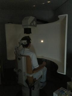
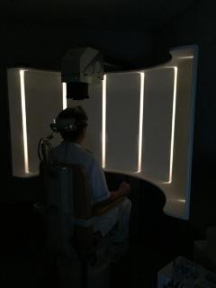
Please sit in front of something like a large screen. Since the white line projected in front of you moves, this is an inspection that allows you to follow this white line with your eyes. Record the eye movement that occurs when the entire field of view is moving. A little after the test starts, you may have the illusion that you are moving. Eye movement that occurs when looking at the outside scenery from a bus or train. The cerebral and cerebellar are involved in the movement of reflective movements that reduce the blur of the eye mark when the head is moving, and the cerebrum and cerebellar that constantly seek to capture the moving eye mark at the center of the retina.
Postoperative eye oscillation test
It is an ocular movement released after visual stimulation. It records ocular vibrations that are said to reflect the function of the brain.
It is said that it reflects the function of the brain that stores eye movement, and if there is a brain disorder, this mechanism is affected and appears in the movement of eye movement.
Inquiries to this page
Yokohama City Apolexy and Spinal Nerves Center
Phone: 045-753-2500 (Representative)
Phone: 045-753-2500 (Representative)
Fax: 045-753-2894
Page ID: 492-297-342


