The text is from here.
Ultrasound
Last updated on April 1, 2022.
- Transcranial ultrasound
- Carotid artery ultrasound
- Ultrasonography of tracheal wall
- Transesophageal heart ultrasonography
- Abdominal ultrasonography
- Lower limb deep vein ultrasonography
Transcranial ultrasound
Transcranial ultrasonography - TC-CFI: transcranial color flow imaging / TCD: transcranial Doppler
Introduction
This is a test to observe the running and blood flow of blood vessels in the skuranial.
Purpose
It is used to evaluate the stenosis and obstruction of blood vessels in the skuranial, and to observe the progress of surgery in the skuranial blood vessels.
In addition, the blood flow in the cranial can be monitored continuously for 30 minutes to search for the presence or absence of micrococks (small grains that clog blood vessels).
Attention
There is no particular pain or pain during the examination. Please wash the bathroom (up to 90 minutes) as the inspection time may be longer in some cases. Inspection is done by applying jelly to the vicinity of the temple or the back of the head, and applying a probe (like a hand scanner).
Some tests may be performed by attaching a fixture to the head to perform monitoring for a long time.
Testing may not be possible depending on the condition of the head bone. There are many difficult cases, especially for older women. The inability to test is not an abnormality, but just the condition of the bone.
There are no dietary restrictions at the time of inspection. You can also take the medicine you usually take.
 State of inspection
 Inspection equipment
Carotid artery ultrasound
Introduction
Cerebral infarction is not a disease caused only by blood vessels in the head. It is often caused by blood vessels from the heart to the head.
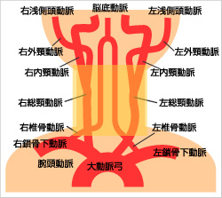
At the neck, there is a blood vessel called carotid artery, which sends blood from the heart to the head. This test observes the blood flow of this carotid artery and the inside of blood vessels.
Purpose

Carotid artery ultrasonography is a test that can evaluate the blood vessels shown in the left figure (total carotid artery: CCA, internal carotid artery: ICA, external carotid artery: ECA, vertebral artery: VA). As shown in the picture below, the vascular cavity can be observed in detail, and the cervical vascular lesions can be observed directly and the presence or absence of arteriosclerosis can be examined. It is also possible to estimate the lesions in distant or near areas by obtaining blood flow waveforms.
Adaptation
Used to evaluate arteriosclerosis changes caused by lifestyle-related diseases and cerebrovascular disorders.
lifestyle-related diseases also contains diabetes, hypertension, hyperlipidemia, stress, insomnia, and smoking. The inspection time is about 30 to 45 minutes.
Attention
There is no particular pain or pain during the examination. The test is performed on the back.
The test is done by applying jelly to the neck and applying a probe (like a hand scanner). Please do not sleep as much as possible during the examination.
As you look at the neck, please come in clothes that can open your neck widely, such as front-armed clothes and V-neck clothes.
There are no dietary restrictions at the time of inspection. You can also take the medicine you usually take.
Inspections use sound reflections, so if the snoring is large, you may get up.
State of inspection
 The appearance of actually applying a probe
 State inside the blood vessels obtained by actually applying a probe
Ultrasonography of tracheal wall
Purpose
Cerebral infarction can be caused by the heart. For example, blood clots formed by depression in the heart blood flow due to myocardial infarction or the spread of left atrium due to arrhythmia, or intra heart tumors.
For this reason, a heart test is a very important test to search for the cause of cerebral infarction. The treatment also changes depending on whether the mental function is maintained. Or, in people with high blood pressure or valvular disease, "heart hypertrophy" is often seen, and as it progresses, it causes a decrease in cardio function.
Cardiac ultrasonography is not just a test for cause search. Checking the heart function determines the possible range of exercise for the patient, which is an important test for patients with chronic stroke.
Method
Observe the heart above the chest (from the left edge of the chest to the axillary through the gaps between the ribs. We observe using ultrasonic waves while applying jelly and applying a probe (like a hand scanner). It is one of the tests commonly referred to as "echo".
Attention
You will take off your jacket and check it with your upper body naked, so it is desirable to wear clothes that are easy to take off and wear.
We may ask you to limit your diet.
There are no restrictions on medications. There are no special precautions in other cases.
Transesophageal heart ultrasonography
Purpose

Probe
Cerebral infarction can be caused by the heart. For example, blood clots formed by depression in the heart blood flow due to myocardial infarction or the spread of left atrium due to arrhythmia, or intra heart tumors.
A normal cardiac ultrasonography (a test that is observed on the chest) is a "examination of the heart from the front", but an esophageal heart ultrasonography observes the heart from the back.
As a result, it is possible to observe parts that are not visible with normal cardiac ultrasonography or to observe them in more detail.
Method
There is an esophagus on the back of the heart. In transesophageal heart ultrasonography, a tube like a gastric camera is swallowed and observed from this esophagus side, that is, the heart from the back side. The procedure of the examination is almost the same as the gastric camera.
Like a gastroscope, first eliminates the vomiting reflex (reflection that tries to exhale with "Oy") and makes the throat anesthesia so that the tube can be swallowed smoothly. For throat anesthesia, use medicine like syrup and spray medicine. You may have to take a deep breath or stop breathing on the way.
This will be done by one doctor and one laboratory technician.
Attention
As you may know if you have received a gastric camera, you can't eat or drink during that time because the throat anesthesia lasts for about 2 hours.
Precautions before the test
It may be accompanied by vomiting reflex when swallowing the tube. If you have any remaining stomach contents, you will be vomiting. For this reason, meals should be kept at least 6 hours before the test and water should be kept at least 4 hours before the test.
If you have a denture, please remove it before the examination.
Don't drive your own car.
Precautions after inspection
Since it uses a medicine that makes it a little sleepy, it may be drowsiness or fluff after the examination.
After the examination until drowsiness, fluffiness, etc. disappear, please return after taking a sufficient rest in the outpatient treatment room. Also, please do not drive your car when you return.
A sore throat anesthesia is performed during the test. If throat anesthesia is effective, things may not be swallowed well, and may accidentally enter the bronchial and cause pneumonia. Sore anesthesia lasts for about 2 hours, so do not eat or drink during that time, and saliva and sputum should be exhaled.
Two hours later, if you drink a little water and swallow it without smash, you can usually eat drinks and meals after that. If you're still getting rid of it, look at it again for about 30 minutes.
Abdominal ultrasonography
Purpose
Abdominal ultrasonography searches for the presence of organic diseases in the abdominal organs (liver, gallbladder, kidney, pancreas, spleen, etc.).
Stroke is common in the elderly, so liver and renal function can be reduced, and drug-related liver dysfunction can also occur.
Stroke has also been associated with lifestyle-related diseases (hypertension, hyperlipidemia, diabetes) and drinking and smoking habits. For this reason, evaluation of the liver etc. is also important.
Method
At the time of the test, lie on your back, apply jelly on the abdomen, and observe using ultrasound while applying a probe (like a hand scanner). It is one of the tests commonly referred to as "echo". As a result, the morphology of each organ is diagnosed with fault images.
Attention
Please come in a fast state during the inspection. In the morning, please complete the meal by 9:00 pm the day before, and in the afternoon inspection by 8:00 am on the day.
As for drinks, small amounts of water and tea may be used, but please avoid milk.
At the time of the examination, the abdomen will be exposed, but it is enough to roll up without undressing. Please come in clothes that are easy to take off or wear, or wear clothes that are easy to roll up.
Lower limb deep vein ultrasonography
Purpose
Cerebral infarction is not a disease caused only by blood vessels in the head. It is often caused by blood clots formed in the veins, especially in the lower limbs.
Blood clots formed in the left centrifugal system or arteries of the heart can block the cerebrovascular vascular, but blood clots formed in the right centrifugal system or in the veins are captured by blood vessels in the lungs returning to the left centrifugal system and enter the arteries. It does not reach the cerebrovascular vascular and does not cause embolism and becomes embolism.
However, if there is a gap in the wall that separates the left centrifugal system of the heart from the right centrifugal system, blood clots formed in the right centrifugal system or veins may reach the cerebrovascular and cause cerebral infarction. This is called "mutant cerebral embolism."
It is said that venous blood clots consist of fibrines, red blood cells, platelets, and white blood cells, and blood clots often occur in areas where blood flow is stagnant near veins and lower thighs. If you sit and stand for a long time, the blood flow of the lower limb veins is depressed and a blood clot is formed.
This test involves examining blood vessels in the deep veins of the lower limbs, blood flow, and checking for blood clots.
Method
It is one of the tests commonly called "echo", and the observation site is from the base of the foot to the top of the knee in the supine position. Apply jelly to the observation site and observe with ultrasonic waves while applying a probe (like a hand scanner). Next, check the veins between the ankle and knees in a sitting position on the bed.
Lower your pants, place a probe at the base of your lower limbs, and repeatedly search for compressions to the top of your knees. This means that if there is a blood clot in the blood vessel, the blood vessels will not be flattened even when compressed. In addition, he sucks large, breaths and repeats exhales several times, and squeezes the calf of the subject.
Attention
The lower limbs are tested by applying jelly. Please come in clothes that are easy to take off or wear, or wear clothes that are easy to roll up.
Inquiries to this page
Yokohama City Apolexy and Spinal Nerves Center
Phone: 045-753-2500 (Representative)
Phone: 045-753-2500 (Representative)
Fax: 045-753-2894
Page ID: 952-614-502


